
Figure 6.
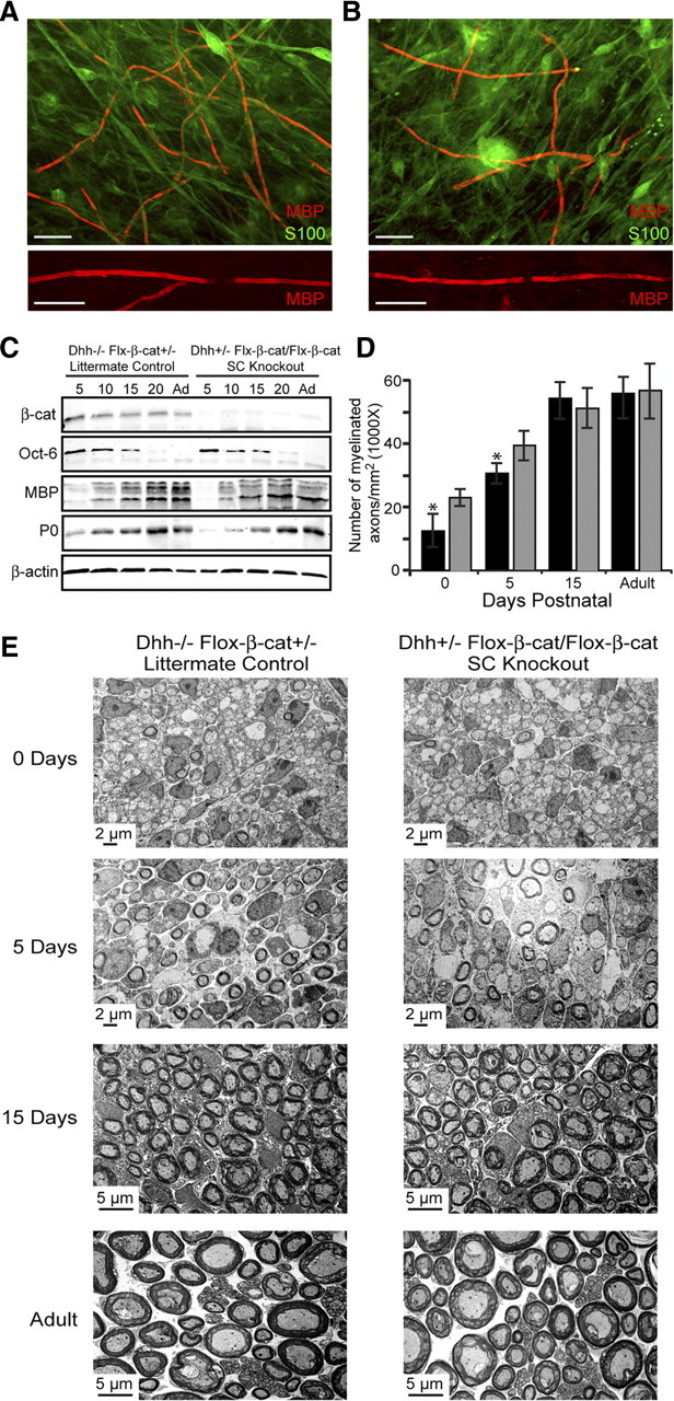
β-Catenin knock-out mice display a more severe delay in myelination without impacting SC numbers, ensheathment, myelin structure, and compaction. A, B, SC–DRG cocultures were established from SC-specific β-catenin knock-out mice (B) (Dhh-Cre+/−, Flox-β-cat/Flox-β-cat) and littermate controls (A) (Dhh-Cre−/−, Flox-β-cat+/−). Myelination was assessed by immunostaining for MBP (red) and S100 (green) 10 d after induction of myelination. The bottom panels illustrate magnified views of myelin internodes formed by SCs from both mice. Scale bar, 20 μm. C, Western blot analysis was performed using mouse sciatic nerves from SC-specific knock-out mice and littermate controls. Westerns were probed for β-catenin, Oct-6, MBP, P0, and β-actin (loading control). D, Myelination in knock-out and control mice was quantified as the number of myelinated axons per square millimeter. The black columns represent the knock-out nerves, and the gray columns are the littermate controls. Nerves from three separate mice were analyzed, and the values are displayed as the mean number of myelinated axons ± SD. Significance was evaluated using Student's t test (*p < 0.05). E, The number of Schwann cells, axonal sorting, compaction, and extent of myelination were examined by electron microscopy of knock-out and control mouse sciatic nerves at 0, 5, 15 d postnatal and adult nerves.