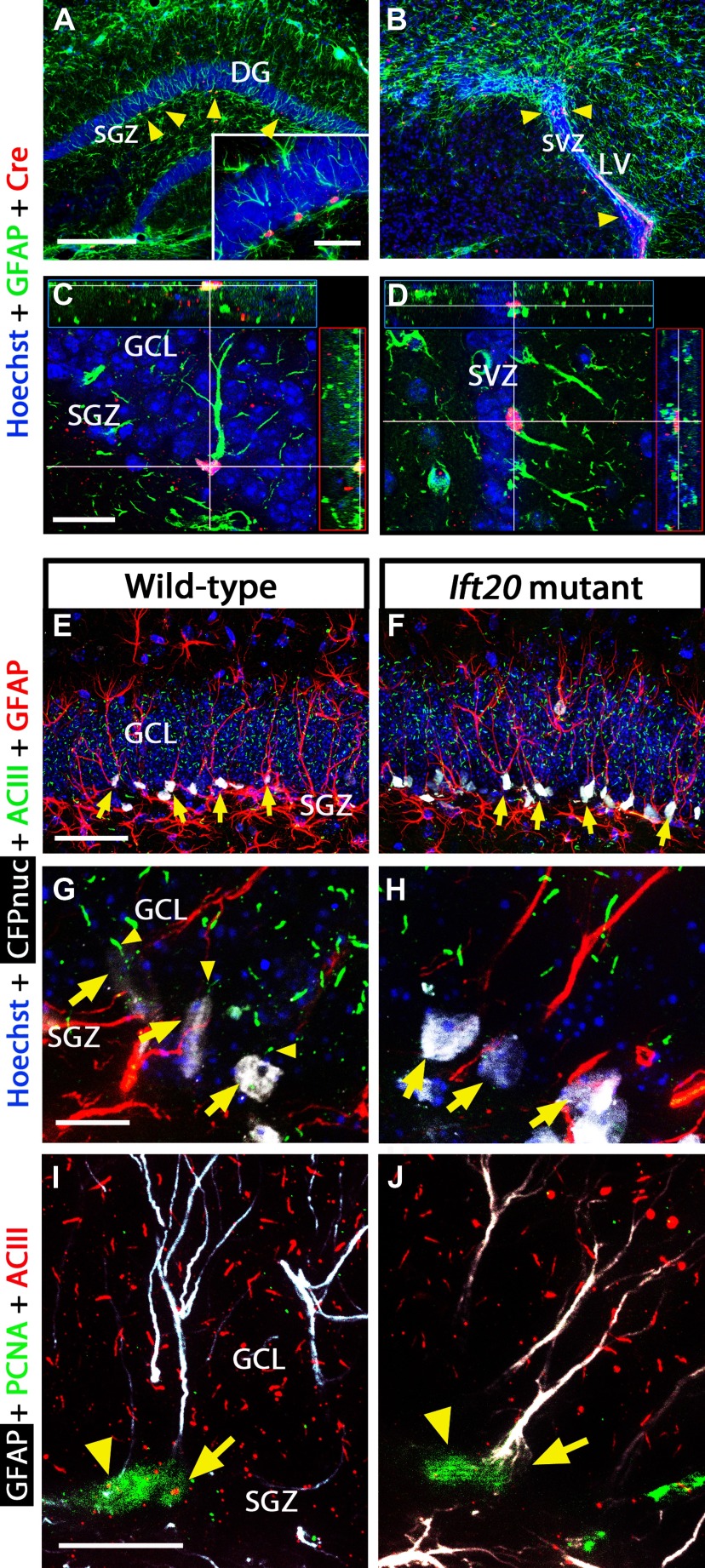
Figure 1.
Primary cilia are ablated in GFAP-expressing NSCs from Ift20fl/fl::mGFAP-Cre (Ift20 mutant) mice (Ift20+/+::mGFAP-Cre; wild type). Double immunostaining in 8-week-old Ift20 mutant mice shows Cre expression (red) in GFAP+ cells (green; arrowheads) in the SGZ (GCL, granule cell layer) of the hippocampal DG (A) and in the SVZ of the LVs (B). Confocal imaging showing GFAP+ cells expressing Cre in the SGZ (C) and in the SVZ (D) of 8-week-old Ift20 mutant mice. Confocal imaging with CFPnuc (white), ACIII (green; marker for primary cilia), and GFAP (red) in the DG showing primary cilia and GFAP-expressing quiescent NSCs (and hence are nestin-CFPnuc+; arrows) from 8-week-old Ift20+/+::mGFAP-Cre::nestin-CFPnuc (wild-type) (E) and Ift20fl/fl::mGFAP-Cre::nestin-CFPnuc (Ift20 mutant) (F) mice. Confocal imaging with CFPnuc (white), ACIII (green), and GFAP (red) in the SGZ of the DG showing primary cilia (arrowheads) in GFAP-expressing radial NSCs (arrows) from 8-week-old Ift20+/+::mGFAP-Cre::nestin-CFPnuc (wild type) (G), whereas equivalent cells do not show primary cilia in Ift20fl/fl::mGFAP-Cre::nestin-CFPnuc (Ift20 mutant) (H) mice. I, J, Confocal GFAP (white), PCNA (green), and ACIII (red) triple staining in DG of 8-week-old Ift20fl/fl::mGFAP-Cre (Ift20 mutant) and wild-type mice. Adult hippocampal stem/progenitor cells in cell cycle (PCNA+) show primary cilia staining (ACIII+) in wild-type but not in Ift20 mutants [arrows, radial NSCs (radial GFAP+); arrowheads, other cells in cell cycle]. Section thickness: A–J, 30 μm. Scale bars: A, B, 200 μm; C, D, I, J, 20 μm; E, F, 50 μm; G, H, 10 μm.

