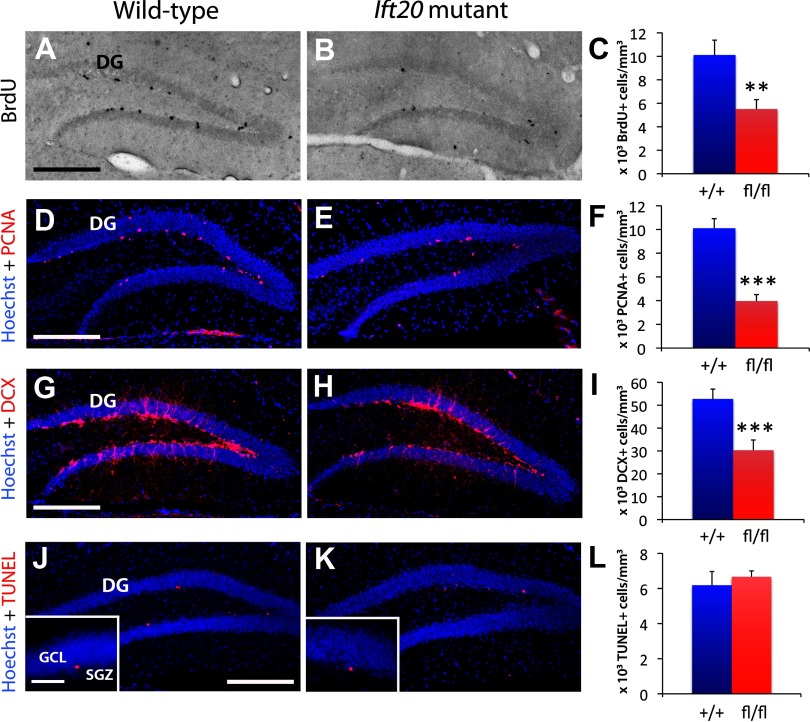
Figure 3.
Ift20 mutant mice exhibit reduced neural precursor cell proliferation in the SGZ of the DG. A, B, BrdU (black) staining after a 2 h BrdU pulse (100 mg kg−1 body weight) showed less proliferation in the SGZ of the DG in 8-week-old Ift20 mutants than in wild-type mice (quantified in C). D, E, Staining with the proliferative marker PCNA (red) confirmed that 8-week-old Ift20 mutants have approximately one-half the number of proliferating cells in the DG as wild-type mice. F, Quantification of PCNA+ cells. G, H, DCX-positive cell population (red) is reduced in the DG of 8-week-old Ift20 mutants. I, Quantification of DCX+ cells. J, K, Apoptosis (red) in the DG, as evaluated using the ApopTag Red In Situ Apoptosis Detection Kit (Millipore Bioscience Research Reagents), was not affected in 8-week-old Ift20 mutants relative to wild-type mice. L, Quantification of apoptotic cells. Section thickness: A, B, D, E, G, H, J, K, 30 μm. Scale bars: A, B, D, E, G, H, J, K, 200 μm; J, K, inset, 50 μm. **p < 0.01; ***p < 0.001. Data are expressed as mean values ± SEM.

