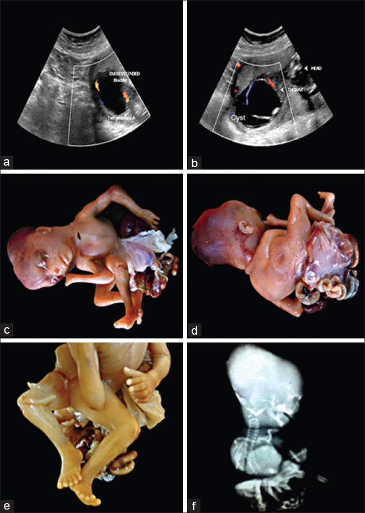
Figure 1.

(a) Ultra sonograms showing a large cyst, in the wall of cyst umbilical vessels are seen (b) Ultra sonogram showing parts of fetus (note the large cyst in the lower part of the fetus) (c and d) Gross photograph of the fetus showing encephalocele, amelia of digits of right upper limb, abdominal wall defects with protrusion of organs which are covered by thin membrane, scoliosis and malrotation of the lower limbs (e) Closer view of lower limbs showing malrotation (lower limbs digits are arranged in opposite direction) (f) Infantogram showing encephalocele and scoliosis
