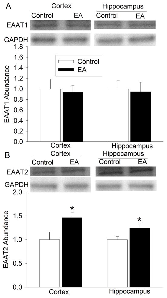
Fig. 5. Effects of electroacupuncture on excitatory amino acid transporter (EAAT) expression in EAAT type 3 knockout mice.
Cerebral cortex and hippocampus were harvested for Western blotting at 24 h after the last electroacupuncture without the MCAO. The EAAT1 and EAAT2 expression is presented in the panels A and B, respectively. Representative Western blots are shown on the top panel and the graphic presentation of the EAAT protein abundance quantified by integrating the volume of autoradiograms from 5 mice for each experimental condition is shown on the bottom panel. Values in graphs are the means ± S.E.M. * P < 0.05 compared with control mice inhaling oxygen 30 min each day for 5 days. GAPDH: glyceraldehydes 3-phosphate dehydrogenase; EA: electroacupuncture.

