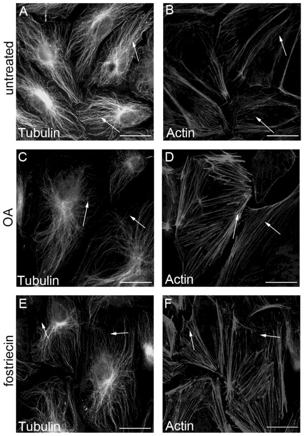
Figure 1. PP2A inhibition affects the organization of cytoskeleton structure.
HLMVEC (A–F) monolayers were treated either with 0.1% DMSO (A,B), with 5 nM OA for 90 min (C,D), or with 100 nM fostriecin for 1 hr (E,F), then the cells were double stained as described in Materials and Methods with anti-β-tubulin primary antibody (A,C,E) and with Texas Red–phalloidin (B,D,F) to visualize the microtubules and microfilaments, respectively. Pictures were taken with an Olympus Fluoview FV1000 confocal microscope, scale bars: 200 μm. A and B, C and D, E and F are parallel images. Shown are representative data of three independent experiments.

