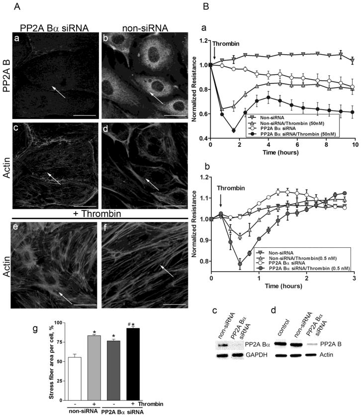
Figure 3. PP2A Bα depletion affects endothelial cytoskeleton organization and barrier function.
Cells were transfected with small interfering RNA (siRNA) specific for PP2A Bα or with non-silencing (non-si) RNA. Panel A: Immunofluorescent staining of BPAEC transfected with PP2A Bα specific siRNA (a,c,e) and non-silencing siRNA (b,d,f). Transfected monolayers without any further treatment were double-stained with PP2A B specific antibody (a,b) and with Texas Red-phalloidin (c,d). Actin staining of transfected cells challenged with thrombin (50 nM, 30 min) (e,f) is also shown. Stress fiber formation induced by thrombin was evaluated by morphometric analysis using Image J program as described in Materials and Methods (g). The results are presented as means n=14, ± SEM. Significant changes are indicated by * (P<0.05 vs control), and # (P<0.05 vs non-si/thrombin). Pictures were taken with a Zeiss Axiolab microscope, scale bars: 200 μm. Panel B: HPAEC (a) and HLMVEC (b) plated on gold microelectrodes were transfected with non-targeting or specific PP2A Bα siRNA. 72 hours later TER was measured. Arrow indicates the time point when thrombin (a:50 nM or b:0.5 nM) or vehicle was added to the medium. The depletion was verified by RT-PCR (Bc) using specific primer pairs; and by Western blot (Bd) of the lysate of control cells and lysates of cells transfected with non-si or PP2A B specific siRNA using B subunit specific antibody. GAPDH and actin signals were used as inner and loading controls, respectively. RT-PCR and Western blot verification was done for all cell types investigated. The efficiency of depletion was about the same, results shown (Bc-d) were acquired on HLMVEC.
Shown are representative data of three independent experiments.

