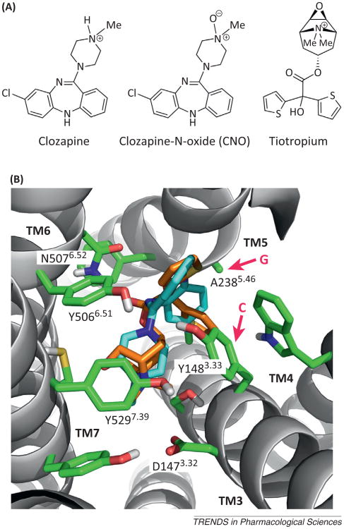
Figure 2.
Docking of clozapine-N-oxide (CNO) into the ligand binding pocket of the rat M3 muscarinic receptor (M3R). (A) Chemical structures of clozapine, CNO, and tiotropium. Note that CNO differs from clozapine only in the presence of the N-oxide moiety. The structure of tiotropium, a muscarinic antagonist/inverse agonist, is shown for comparison because of the availability of a high-resolution X-ray structure of the tiotropium–rat M3R complex [17]. (B) Extracellular view of the tiotropium binding pocket [17]. Key amino acids that are of particular importance for tiotropium binding are highlighted. Both Y3.33 and A5.46 (red arrows) are predicted to contact the tiotropium ligand (see the text for details; note that all muscarinic receptor-based DREADDs contain the Y3.33C and A5.46G point mutations). The amino acids that replace Y3.33 and A5.46 in the DREADD constructs are indicated (C and G at the beginning of the red arrows). CNO can be docked into the M3R binding pocket in a pose very similar to that of tiotropium, without any obvious structural clashes (the two compounds are similar in size and overall shape; CNO, light blue; tiotropium, orange; Dahlia R. Weiss and Brian K. Shoichet, personal communication).

