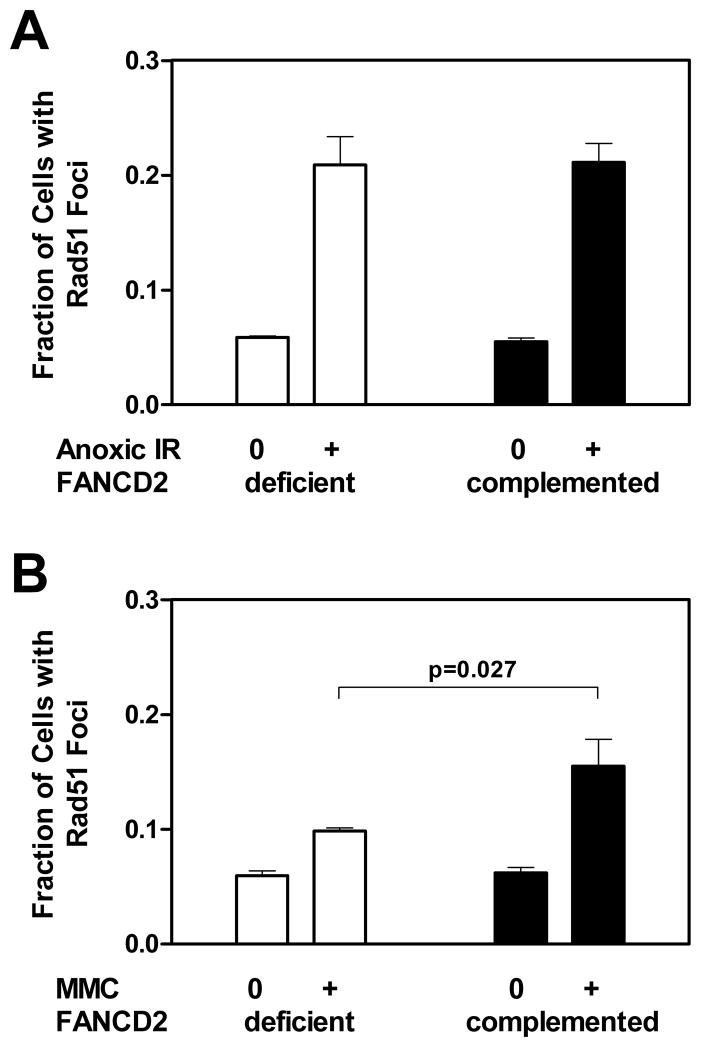
Figure 3.
Formation of RAD51 foci in FANCD2-deficient (PD20) and wild-type complemented (PD20-wtD2) fibroblasts. (A) The fraction of cells with at least 5 foci is plotted for untreated cells (0) versus cells exposed to 16 Gy ionizing radiation (IR) at 0% oxygen (+) after 5 hours. The relative induction of foci was 3.6-fold for FANCD2-deficient cells. (B) Foci formation analogous to panel A 5 hours following mock-treatment (0) or treatment with mitomycin C (MMC) at 0.5 μg/ml (+). The induction was 1.7-fold for FANCD2-deficient cells. The relative induction of foci above untreated levels between the two cell lines was compared using the unpaired t-test (two-sided p-value). Bars represent means with standard error based on three independent repeat experiments.

