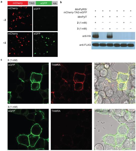
Figure 4. Site-specific incorporation of 2 into proteins in mammalian cells and the specific labelling of EGFR-GFP on the cell surface with 9.
a, Cells that contain the PylRS/tRNACUA pair and the mCherry(TAG)eGFP-HA reporter produced GFP only in the presence of 2. b, Western blots confirm that the expression of full length mCherry(TAG)eGFP-HA is dependent on the presence of 2. c, Specific and rapid labelling of a cell surface protein in live mammalian cells. EGFR-GFP that bears 2 or 3 at position 128 is visible as green fluorescence at the membrane of transfected cells (left panels). Treatment of cells with 9 (200 nM) leads to selective labelling of EGFR that contains 2 (middle panels). Right panels show merged green and red fluorescence images, DIC=differential interference contrast. Cells were imaged four hours after the addition of 9.

