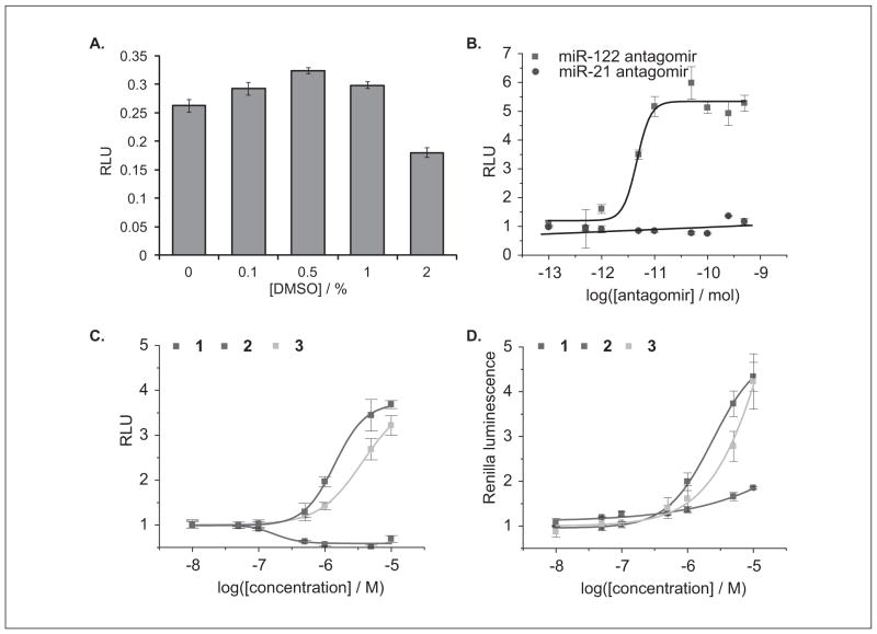
Figure 4.
(A) Relative luminescence units (RLU) of Huh7-psiCHECK-miR122 cells treated with increasing concentrations of DMSO. All assays were conducted in triplicate in a 96-well format. (B) Luciferase assay dose-response curves for Huh7-psiCHECK-miR122 cells transfected with either an miR-122 or miR-21 antagomir in a 96-well format. All assays were conducted in triplicate and were normalized to a control containing only transfection reagent. (C) Dose-dependent response of RLU for Huh7-psiCHECK-miR122 cells treated with increasing concentrations of the miR-122 inhibitors 1-3. All assays were conducted in triplicate in a 384-well format and were normalized to a DMSO control. (D) Dose-dependent response of absolute Renilla luminescence units for Huh7-psiCHECK-miR122 cells treated with increasing concentrations of the miR-122 inhibitors 1-3. All assays were conducted in triplicate in a 384-well format and were normalized to a DMSO control. Error bars represent standard deviations from three independent experiments.

