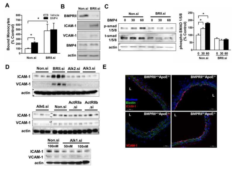
Figure 1. Loss of BMPRII induces endothelial inflammation in vitro and in vivo.
(A, B, C) HUVECs were transfected with BMPRII siRNA (BRII.si) or non-silencing control siRNA (Non.si). In (A), cells were then treated with BMP4 (100 ng/ml, 4 h), and THP1 monocyte adhesion to ECs was determined (mean ± SEM, n=6, *p < 0.05). In (B), cells were lysed and analyzed by Western blot with antibodies to BMPRII, ICAM-1 and VCAM-1, using β-actin as a loading control. In (C), cells were treated with BMP4 and cell lysates were analyzed with phospho-smad1/5/8 or total smad1/5/8 antibodies, and the Western band intensities were quantified (*p<0.05, n=3). (D) HAECs transfected with siRNAs specific for BMPRII, Alk1, Alk2, Alk3, Alk6, ActRIIa, ActRIIb or control (Non.si) were analyzed by Western blots with BMPRII, VCAM-1 and ICAM-1 antibodies. (E) Representative images show immunostaining of frozen sections of thoracic aortas obtained from BMPRII+/−ApoE−/− and BMPRII+/+ApoE−/− mice (n=6 each) using ICAM-1 and VCAM-1 antibodies (red). Nuclei stained with DAPI (blue) and auto fluorescence signals showing elastic laminas are shown in green. L: lumen.

