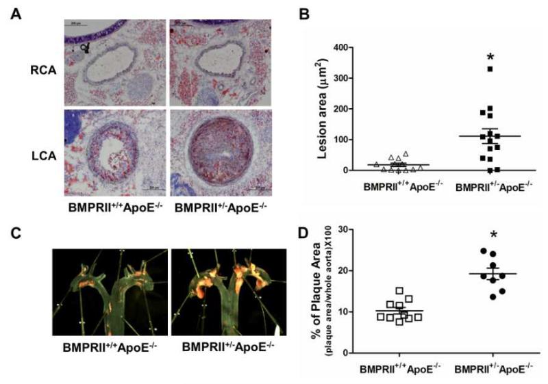
Figure 3. BMPRII deficiency exacerbates atherosclerosis development in ApoE−/− mice.
(A) BMPRII+/−ApoE−/− (n=12) and littermate control BMPRII+/+ApoE−/− mice (n=14) were partially ligated and fed a high-fat diet for 2 weeks. Frozen sections from the ligated LCA (d-flow) and contralateral RCA (s-flow) were stained with Oil-Red-O as shown by representative microscopy images. (B) Atherosclerotic lesion area was quantified (*p<0.001). (C) BMPRII+/−ApoE−/− (n=8) and BMPRII+/+ApoE−/− mice (n=10) were fed the high-fat diet for 2 months. Aortic arch regions were stained with Oil-Red-O as shown, and (D) atherosclerotic lesion areas determined (*p<0.05).

