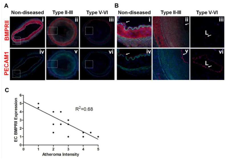
Figure 5. Loss of BMPRII in human coronary arteries with advanced atherosclerotic plaques.
(A, B) Representative confocal microscopy images of human coronary arteries containing various stages of atherosclerotic lesions were stained with antibodies to BMPRII (i-iii) and PECAM-1 (iv-vi), shown in red. DAPI (blue); auto fluorescence matrix signals (green). Images shown in (B) are magnified regions indicated by broken boxes in (A). (C) The graph shows semi-quantification of BMPRII staining intensity in the endothelial layer as a function of atheroma severity carried out by two blinded investigators (n=14).

