
Fig. 2.
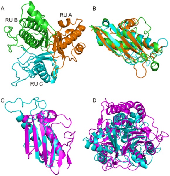
Structure of AtzD monomer and comparison to the perchloric acid soluble protein (PSP).
A. Structure and substructure of an AtzD monomer. The RUs are coloured as follows: RU A (residues 2–104) is orange, RU B (residues 113–250) is green and RU C (residues 256–364) is cyan.
B. Overlay between RUs A, B and C (coloured as Fig. 2A).
C. PSP (PDB: 3K0T) monomer (magenta) superposed with RU C of AtzD (cyan).
D. A trimer of PSP (magenta) superposed with a monomer of AtzD (cyan).