
Fig. 3.
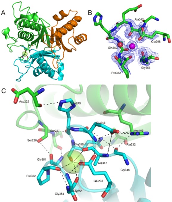
The metal binding site of AtzD.
A. The position of the bound metal (green sphere) in the AtzD monomer (coloured as per Fig. 2A).
B. The bound metal (magenta sphere), the metal-binding residues, the water ligand (red sphere) and their electron densities are shown.
C. The interactions between the metal, metal-stabilized loop and the surrounding protein.