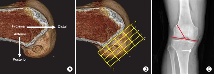
Fig. 1.

(A) The medial-lateral view of the lateral femoral condyle is obtained from the 3-dimensional computed tomography, and is rotated to the strictly lateral position. (B) A rectangular measurement frame is drawn over the medial-lateral view of the lateral femoral condyle described by Bernard et al.8) (C) Lateral inclination of the femoral bone tunnel was measured with reference to a line tangent to the femoral condyle.18)
