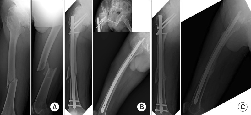
Fig. 2.
(A) Initial anteroposterior and lateral views show Arbeitsgemeinschaft für Osteosynthesefragen type B1 femoral shaft fracture. (B) Immediate postoperative anteroposterior, lateral and oblique views show additional fracture in the lateral cortex of the proximal femur. (C) Postoperative 1 year radiographs show delayed union in the femoral shaft.

