
Figure 1.
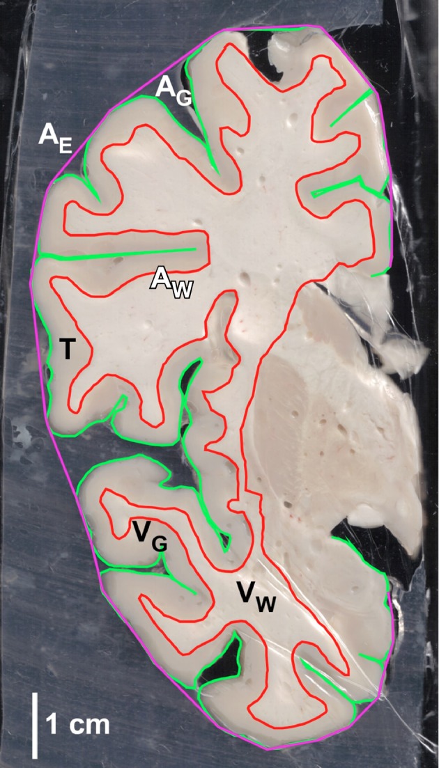
Example of coronal section of the human cerebral cortex with reconstructions of outlines for determination of total cortical area (AG), exposed cortical area (AE), white-gray matter interface area (Aw), gray matter volume (VG) and white matter volume (VW), and gray matter thickness (T).