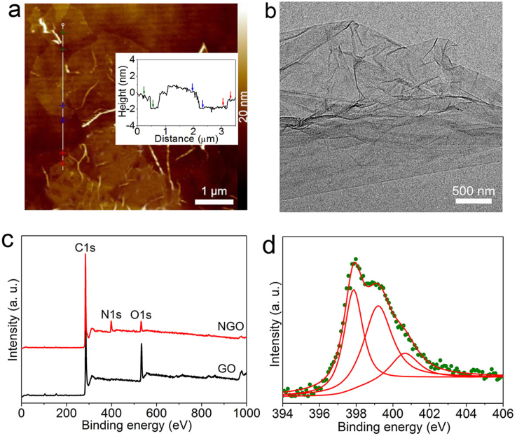
Figure 1. Microstructure of GO and NGO.
(a) Tapping mode AFM height image of GO. Inset: The corresponding height profiles of the white lines with colorful crosses. The colorful arrows correnspond to the colorful crosses. (b) TEM image of GO. (c) XPS spectra of GO and NGO over a wide range of binding energies (0–1000 eV). (d) Fine-scanned N 1s XPS spectrum of NGO. The olive dots and red lines are measured dots and fitting curves.

