Figure 4.

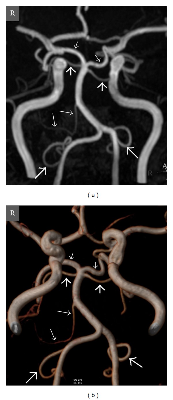
Oblique coronal view MIP (a) and VR (b) TOF MR angiography images show absence of the left AICA, well-developed left PICA, and moderately developed right PICA in a 48-year-old woman. Thick long arrows = right and left PICAs, thin long arrow = right AICA, thick short arrows = right and left SCAs, thin short arrows = right and left PCAs, and R = right.
