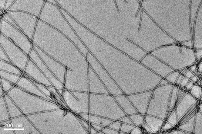
Figure 2. TEM micrograph of I3K nanofibrils.

The peptide solution (4 mM in water) was incubated for 1 week at pH of around 7 and the sample was negatively stained with 2% uranyl acetate before TEM imaging.

The peptide solution (4 mM in water) was incubated for 1 week at pH of around 7 and the sample was negatively stained with 2% uranyl acetate before TEM imaging.