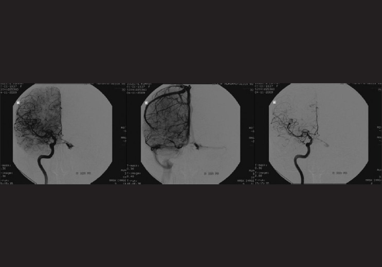
Figure 3.

Cerebral angiography shows a dural carotid cavernous fistula on the right side draining through the right superior opthalmic vein as well as the left superior opthalmic vein via posterior intercavernous sinus connection (arterial and venous phase)
