Figure 1.

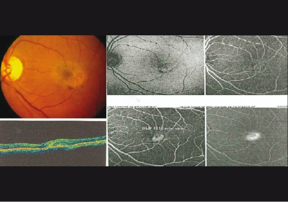
Left eye fundus photograph showing stage 5 idiopathic macular telangiectasia (IMT) confirmed on optical coherence tomogram (OCT) and fundus fluorescein angiography (FFA)

Left eye fundus photograph showing stage 5 idiopathic macular telangiectasia (IMT) confirmed on optical coherence tomogram (OCT) and fundus fluorescein angiography (FFA)