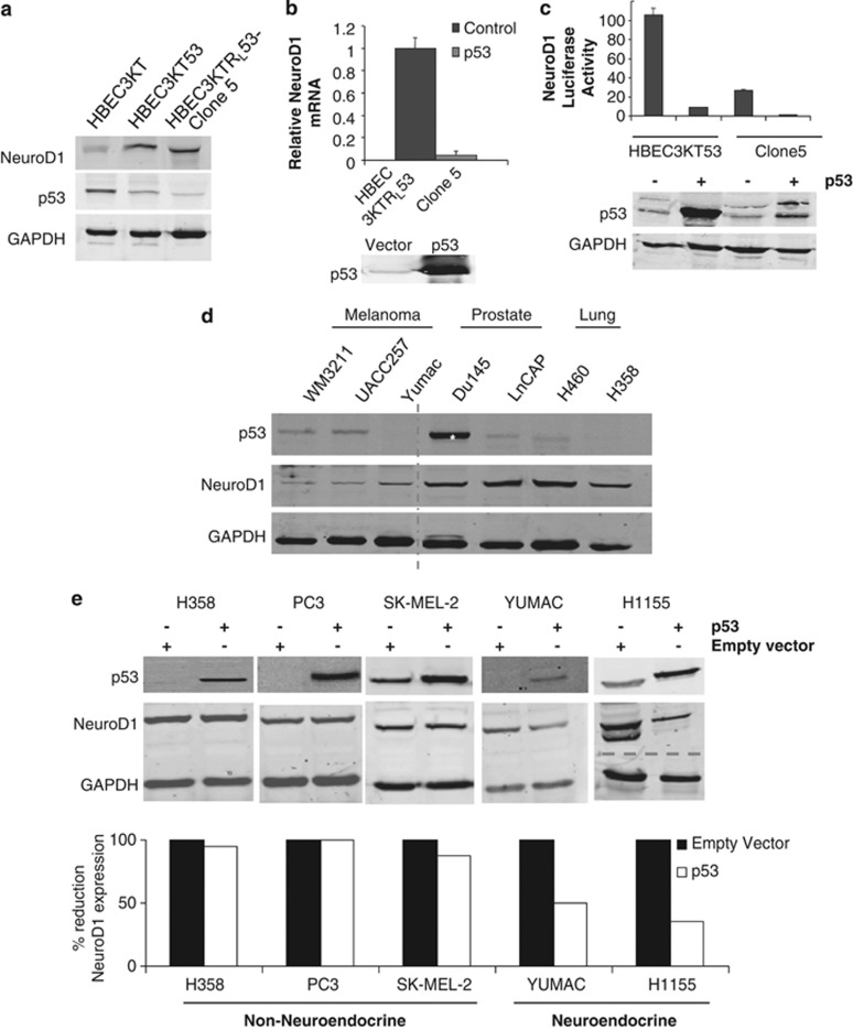
Figure 2.
Loss of p53 is permissive for expression of NeuroD1. (a) NeuroD1, p53 and GAPDH (loading control) were immunoblotted in lysates of HBEC3KT, HBEC3KT53 and Clone 5. (b) qRT–PCR analysis of NeuroD1 in Clone 5 cells transfected as indicated. A representative p53 immunoblot is shown from one of three independent experiments. (c) HBEC3KT53 and Clone 5 were transfected with pGL3-NeuroD1-luciferase with and without p53. p53 was immunoblotted and luciferase activity was measured; one of six experiments shown. (d) Melanoma, prostate and lung cancer cell lines were lysed; 50 μg total protein was immunoblotted for p53, NeuroD1 and GAPDH (as loading control). The dashed line indicates discontinuity in gel. The asterisk represents a loss-of-function mutation in p53.43 (e) Cell lines with loss of or mutation in p53 were transfected with control vector or vector encoding p53. Cells were lysed and immunoblotted for p53. Overexpression was quantified using Odyssey software.

