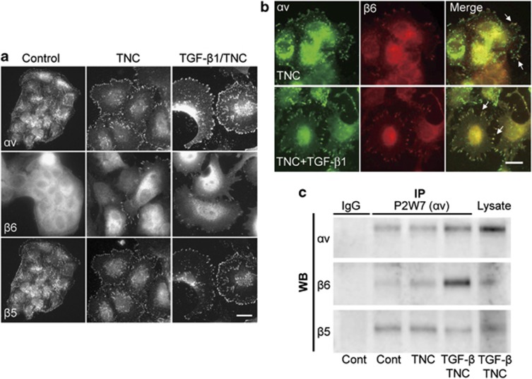
Figure 3.
Integrin β6 subunits are recruited to αv-positive adhesion plaques. (a) Immunofluorescence showed control cells to express αv (P2W7) and β5 integrin subunits, but not the β6 subunit. After TNC only and the TGF-β1/TNC treatment, β6-positive adhesion plaques were observed, more frequently in TGF-β1/TNC-treated cells. (b) Double immunofluorescence staining demonstrating colocalization of αv (Q-20) and β6 subunits. Scale bars: 20 μm. (c) Immunoprecipitation with anti-αv antibody (P2W7) showing prominent increase of co-precipitated β6 subunit in TGF-β1/TNC-treated cells. SDS-gel electrophoresis was performed under non-reducing conditions. As a negative control, a monoclonal antibody against a viral protein was used instead of anti-integrin antibodies. Whole-cell lysates (Lysate) of TGF-β1/TNC-treated cells were also examined.

