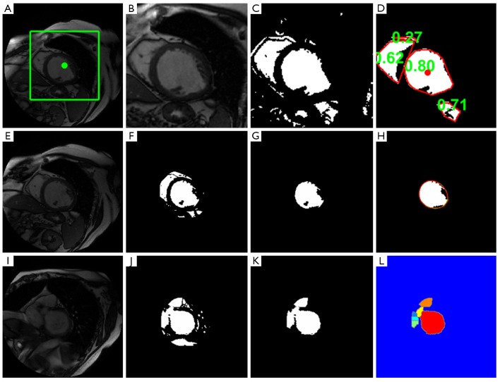
Figure 1.
Left ventricle (LV) localization, endocardial contour detection and outflow tract segmentation. A-D. LV localization procedure; E-H. LV endocardial contour detection; I-L. Identify and segment basal slice with LV outflow tract. A. Target image with rectangular region-of-interest (ROI) and image center, (green square and point); B. ROI image; C. Binary image; D. Surviving objects’ convex hulls (red), the corresponding roundness metrics (green), and the detected LV blood pool centroid (red point); E. Target image; F. Binary image masked by ROI. G. Located LV blood pool; H. Smoothed endocardial contour (red), and papillary muscles and trabeculations (black regions in the smoothed endocardial contour); I. Basal slice image with LV outflow tract (LVOT); J. Binary image masked by ROI; K. Blood pool including LV; L. Watershed results, Watershed results, with detected LV blood pool (red), LVOT (yellow), right ventricle outflow tract (orange). Other colored objects indicate regions from right ventricle and atrium

