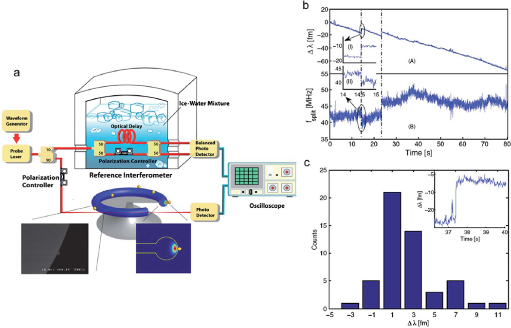
Fig. 8.
High Sensitivity Optical Microcavity based detector. (a) Experimental setup for nanoparticle detection using a temperature-stabilized reference interferometer. The output of a tunable laser is split into two branches by a 90/10 coupler. One branch is coupled into/out of a microtoroid resonator in a cooled aqueous environment. The other branch is coupled into a reference interferometer to monitor the laser optical frequency in real time. The reference interferometer is immersed in ice-water to improve the stability. (Inset) SEM micrograph of an R = 25 nm bead binding on the surface of a microtoroid. (b) The resonance wavelength shift (scan A) and splitting frequency shift (scan B) are shown for a microtoroid immersed in a 1 pMInfA solution. The insets I and II show that the same resonance wavelength shift event can also be detected as a split frequency shift, respectively. (c) The histogram of the resonance wavelength-shift steps in scan A and an inset of the largest wavelength step recorded of 11.3 fm. Adapted from ref. 99. © 2011 National Academy of Sciences.

