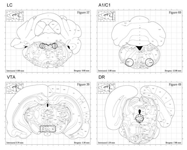
Figure 1.
Brain sections showing representative dissections (punches or cuts) taken to obtain samples of the locus coeruleus (LC), A1/C1 brainstem (A1/C1), ventral tegmental (VTA), and dorsal raphe nucleus (DRN) brain regions. For LC, tissue was obtained by 1.0 mm diameter bilateral punches of three contiguous 1.0 mm thick sections; for A1/C1 and DRN, tissue was obtained by 1.5 mm diameter punches of two contiguous 1.0 mm thick sections on midline (DRN) and bilaterally (A1/C1); for VTA, a rectangular dissection on midline (as shown) was taken from two contiguous 1.0 mm thick sections. Brain sections shown are approximately the midpoint of regions dissected. Illustrations are taken from Paxinos and Watson, 1998.

