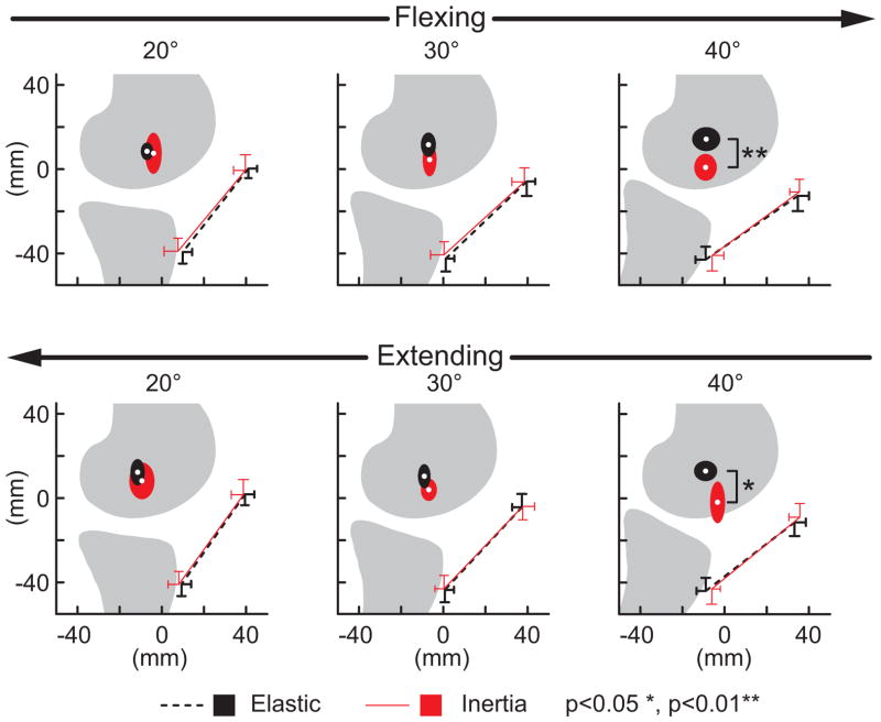
Figure 8.
Shown are the average (±1 sd) origin and insertion of the patellar tendon and the average (±1 sd) location at which the tibiofemoral finite helical axis crossed the mid-sagittal femur. Discrete flexion angles within the flexing and extensing phases of motion are shown. Note the FHA migrated distally with knee flexion when the quadriceps were loaded (inertial case), reducing the moment generating potential of the patellar tendon.

