Figure 4.
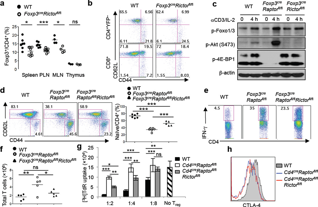
Deletion of Rictor does not alter Treg function but partially rescues inflammation in Foxp3creRaptorfl/fl mice. a, Treg percentage in 2–3 month old wild-type and Foxp3creRictorfl/fl mice. b, Expression of CD62L and CD44 on splenic T cells from wild-type and Foxp3creRictorfl/fl mice. c, Tregs were activated by anti-CD3 and IL-2 for 4 hours followed by immunoblots. d, Expression of CD62L and CD44 expression on splenic CD4+ T cells from 3–4 week old wild-type, Foxp3creRaptorfl/fl and Foxp3creRaptorfl/flRictorfl/fl mice. Right, percentage of CD62LhiCD44lo naïve CD4+ cells in the spleen. e, IFN-γ production in splenic CD4+ T cells from wild-type, Foxp3creRaptorfl/fl and Foxp3creRaptorfl/flRictorfl/fl mice. f, Numbers of total TCRβ+ cells in peripheral lymph nodes from wild-type, Foxp3creRaptorfl/fl and Foxp3creRaptorfl/flRictorfl/fl mice. g, In vitro suppression assays mediated by Tregs from 3–4 week old wild-type, CD4creRaptorfl/fl and CD4creRaptorfl/flRictorfl/fl mice. Error bars represent s.d. (n=3). h, CTLA-4 expression in Tregs from the spleen of wild-type, CD4creRaptorfl/fl and CD4creRaptorfl/flRictorfl/fl mice. P values are determined by Mann Whitney test (a) and ANOVA (d, f, g). Results represent 3 (a-c, e-g) and 2 (d, h) independent experiments.

