
Figure 3.
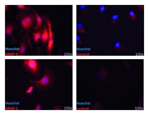
Immunohistochemistry of murine microglia, stained for MMP-2 and MMP-9. Microglia from CD11c GFP mice, fixed with PFA, treated with TBS and NGS, anti-MMP-2 (1 : 125) and anti-MMP-9 (1 : 500); control: BSA and secondary antibodies.
Official websites use .gov
A
.gov website belongs to an official
government organization in the United States.
Secure .gov websites use HTTPS
A lock (
) or https:// means you've safely
connected to the .gov website. Share sensitive
information only on official, secure websites.
Immunohistochemistry of murine microglia, stained for MMP-2 and MMP-9. Microglia from CD11c GFP mice, fixed with PFA, treated with TBS and NGS, anti-MMP-2 (1 : 125) and anti-MMP-9 (1 : 500); control: BSA and secondary antibodies.