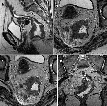
Figure 1. 54-year-old woman with pT4N1 rectal cancer. Two observers staged the tumor as T3N2 in the preoperative MR imaging. a- T2-weighted sagittal MR image shows the tumor. b- T2-weighted para-axial MR image shows the tumor with nodularextramural invasion and perirectal lymph node metastasis. c- On T2-weighted para-axial MR image, the distance betweeninvolved lymph node and mesorectal fascia was more than 1 mm andCRM was defined as uninvolved. But histopathologically CRM wasdefined as involved.d- T2-weighted para-coronal MR image shows the tumor and lymph nodes.

