
Fig. 2.
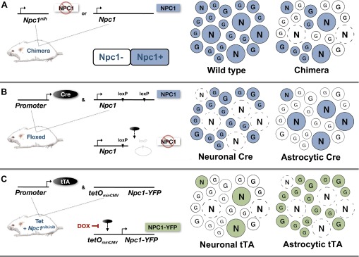
Mouse models of cell-autonomous neuron death and survival in NPC disease. Multiple independent studies using NPC1-deficient mouse models of the disease demonstrate that NPC1 function in neurons is necessary and sufficient to affect neurodegeneration. Mosaic depictions of the various experimental scenarios are shown. N represents neuronal cells and G represents astroglia. Dashed circles represent dead neurons and colored circles are positive for NPC1. (A) Chimeric mice with wild-type and Npc1nih mutant cells provided initial evidence for the cell-autonomous death of neurons in vivo (Ko et al., 2005). In these mice, Npc1+ neurons survived among a mixed Npc1+ and Npc1− glial population but Npc1− neurons did not survive. (B) Npc1 conditional knockout ‘floxed’ mice (in which Npc1 is knocked out by induction of Cre-mediated recombination) were then used to show that deletion of the Npc1 gene in astrocytes does not contribute to neuron loss, whereas deletion of the Npc1 gene in neurons does (Yu et al., 2011). (C) Experiments using NPC1-deficient mice (Npc1nih/nih) engineered for conditional rescue using a Tet-inducible Npc1-YFP transgene further demonstrated cell-autonomous survival of neurons (Lopez et al., 2011). In this study, the subset of neurons that produced NPC1-YFP in an otherwise NPC1-deficient animal survived early disease progression.