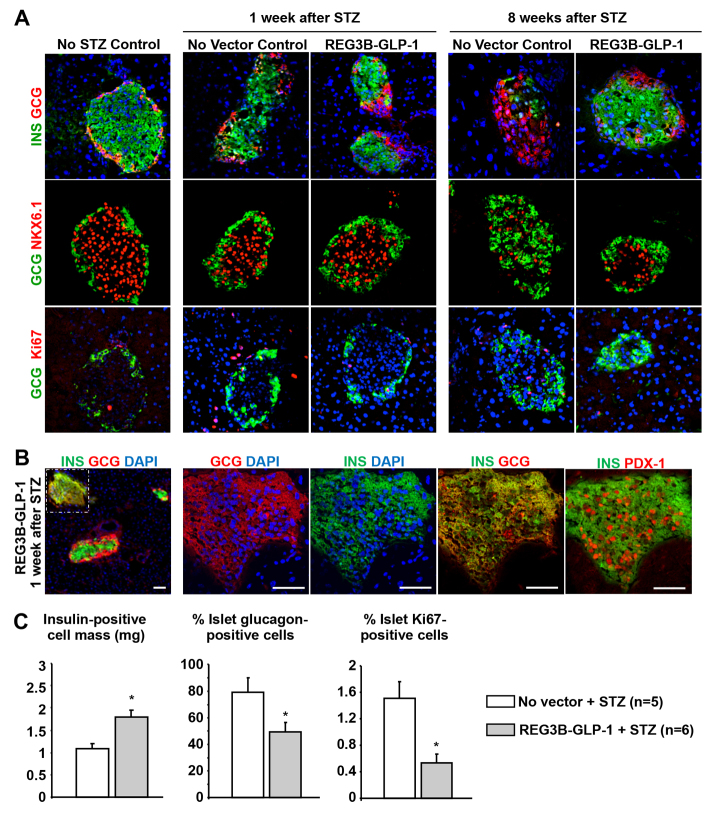
Fig. 3.
REG3B–GLP-1-treated pancreatic islets maintain insulin-positive cells after STZ induction. (A) Immunohistochemistry showing islet structure without STZ and REG3B–GLP-1 (left column), 1-week post STZ induction with or without REG3B–GLP-1 therapy (middle columns), and 8-weeks post STZ induction with or without REG3B–GLP-1 therapy (right columns). Insulin and glucagon (top row), glucagon and Nkx6.1 (middle row), and glucagon and ki67 (bottom row) are shown. All images were taken with 40× objective. (B) Immunostaining of islets 1-week post STZ induction with AAV9 mRIP Reg3b–Glp-1 delivery. A small population of islets was found to be bi-hormonal for glucagon and insulin. The left panel is 10× objective of pancreas cross-section, with the islet with insulin and glucagon dual-positive cells indicated by a dashed box. Other panels show serial sections of a bi-hormonal islet, stained with combinations of anti-insulin, -glucagon or -Pdx1 antibodies, at higher magnifications (40× objective). Nuclei were counterstained by DAPI. Scale bars: 50 μm. (C) 8 weeks after STZ induction insulin-positive cell masses were determined as described in the Materials and Methods (left panel). Percent of total glucagon area to insulin area was determined from five random islets per individual (middle panel). Percent islet cell proliferation was determined by counting the number of ki67-positive cells within insulin- and glucagon-positive regions of five random islets (right panel).

