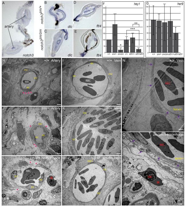
Fig. 7.
Expression of notch3, Notch targets, and arterial and venous markers in adult fin, and analysis of compromised vessel integrity and hemorrhage of vessels in notch3 mutants. (A) Consistent with the vascular phenotypes of notch3 mutants, we detected notch3 expression in association within arteries of adult wild-type (WT) fin. (B) deltaC is expressed in the arteries within the bony segments, but not in the veins, which are located in the joints of notch3zm/+ heterozygotes fins as expected. (C) In notch3zm/zm mutants, deltaC is also expressed in the enlarged vessels (note the enlarged lumen) within bony segments but not in the veins; thus, arterial and venous identity does not require notch3. (D,E) No difference in the pattern of flt4 expression between (D) notch3zm/+ heterozygotes and (E) notch3zm/zm mutant fins was detected, although the mutant vessels were distended. (F,G) qRT-PCR of Notch target expression in caudal fin. (F) qRT-PCR hey1. (G) qRT-PCR her9. y-axis indicates % of expression normalized to WT. (H–O) TEM analysis. (H) Wild-type artery. Pink arrows indicate dense plaques. (I) Wild-type vein. (J) Intact dense plaques surrounded arteries of notch3zm/+ heterozygotes and (K) veins show normal morphology, but appear slightly enlarged. (L) Breaks in the walls and reduced dense plaques (pink arrows) of notch3zm/zm mutant vessels with features reminiscent of arteries. Blood cells (red asterisk) are located outside of the damaged vessel. (M) notch3zm/zm mutant veins are comparable to those of notch3zm/+ heterozygotes. (N) Wild-type vein. The purple arrows in N and O indicate the vessel wall. (O) Hemorrhage and inflammation of notch3zm/zm vessel. Note the red blood cell (red asterisk in O) outside of the compromised vessel wall indicated by purple arrows. Scale bars: 1 μm. ec, endothelial cell; mc, vascular mural cells; RBC, red blood cell. *n=2; **P=0.06; ***P=0.02.

