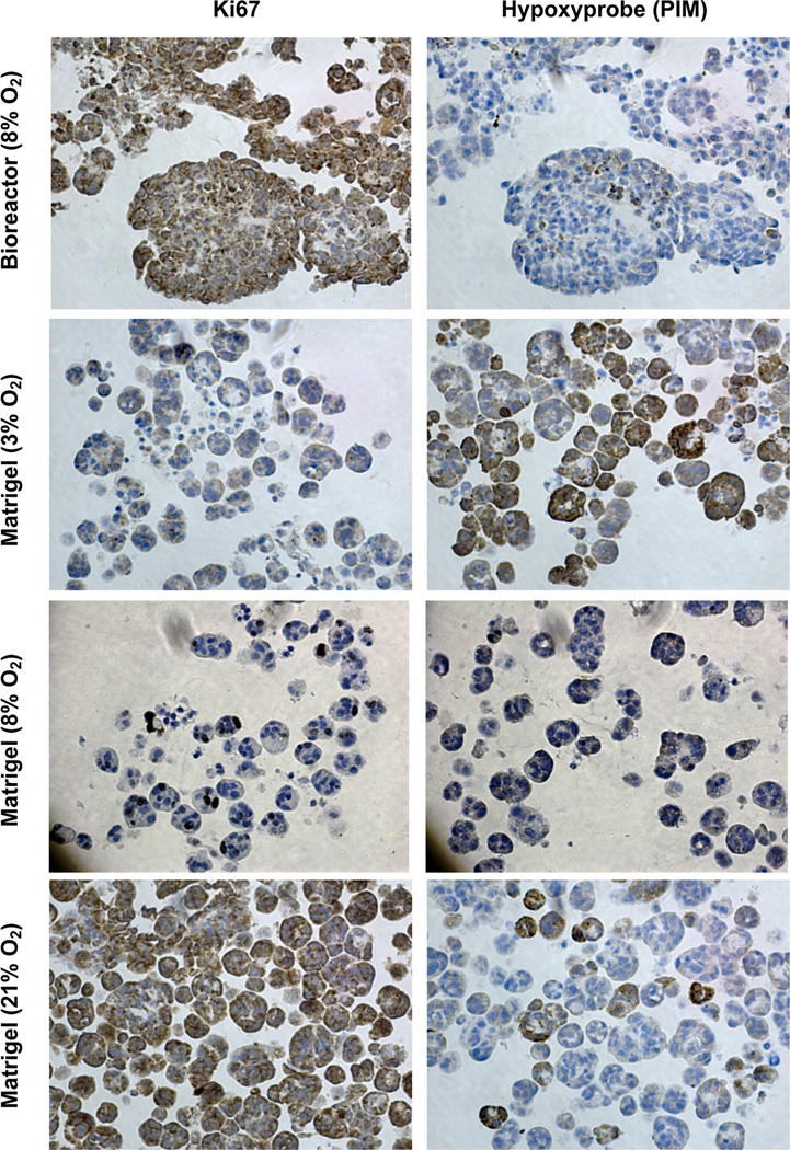
Fig. 7.
Tumor hypoxia and cell proliferation status in 3-D cell culture at different O2 concentrations. OVCAR8-DsRed2 fluorescent cells were cultured for 7 days at 3, 8 and 21% O2 in Matrigel and with 8% O2 in the lower chamber of the bioreactor culture system. Cell proliferation and hypoxia staining were performed with Ki67 for detection of proliferating cells and pimonidazole hydrochloride (Hypoxyprobe) to detect hypoxia within cells, respectively. Increasing O2 concentration showed a corresponding increase in Ki-67 staining and decrease in pimonidazole staining in 3-D Matrigel culture. The bioreactor culture demonstrates a difference in growth morphology compared to 3-D culture in Matrigel and suggests that a lower O2 tension is required to achieve hypoxic culture conditions more akin to in vivo conditions.

