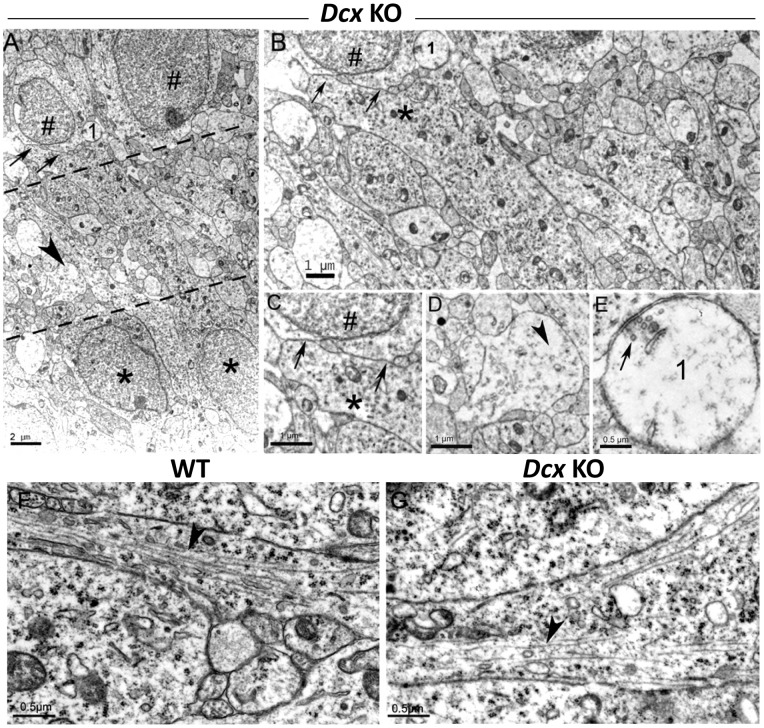
Figure 4. Ultrastructural organization of the intermediary layer (IN).
(A) Low magnification showing the IN delimited by black dashed lines. Cells from the SPI (black symbols #) and SPE (black asterisks) flank numerous neuritic profiles enclosed in the IN layer. The arrows, arrowhead and ‘1’ correspond to zones enlarged in C–E respectively. (B) Higher magnification of the IN. Numerous dense cellular profiles are visualized, corresponding to neuronal processes (likely to be immature dendrites and axons) and synaptic contacts (see ‘E’). A neurite from a cell located in the SPE (black asterisk) extending to and juxtaposing (arrows) the soma from a cell in the SPI (black symbol #). (C) High magnification of the neuropil-like region indicated by two arrows in A and B. Note the close apposition between a probable dendritic profile from an SPE cell and an SPI soma (black symbol #). (D) Higher magnification of arrowhead in A. The cellular profiles of the IN display heterogeneous aspects: some of them with a light cytoplasm correspond to transversal sections of neurites, and contain tubulo-vesicular structures and regions without cytoskeletal elements (arrowhead). (E) is a detail of an axo-somatic synaptic contact identified in A and B as ‘1’. Some synaptic vesicles are observed (arrow). (F, G) Neurites of hippocampal pyramidal neurons. (F) A neurite from a WT pyramidal cell, longitudinally sectioned, contains numerous adjacent microtubules closely arranged in parallel fascicles (arrowhead). (G) In KO, a neurite from an SPI pyramidal neuron shows sparser, more widely-spaced microtubules (arrowhead). Scale bars: A, 2 µm; B, C, D: 1 µm; E–G: 0.5 µm.

