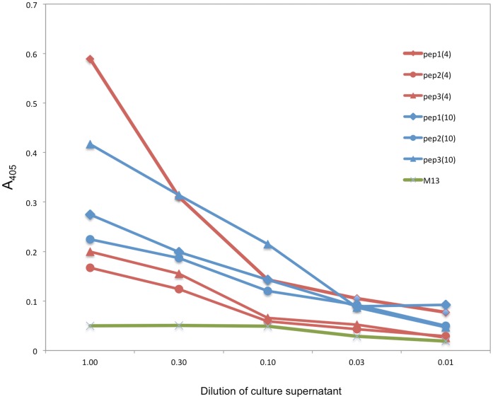
Figure 2. Binding of HCDR3 phage clones to ssDNA.
Individual phage particles derived from chosen V 4 and V
 10 pools were tested. Phage clones with HCDR3 peptides in the context of V
 4 and V
 10 were chosen. Phage supernatant was transferred to oligo-dT adsorbed microplates and serially diluted. Phages were detected with anti-M13 antibodies. Blue, V
 4 phage clones; red, V
 10 clones. Symbols differentiate HCDR3: diamonds, YLLSPLLLA, circle, VQQVNNLA e triangle VQYVNNALA. Green line represents the VCM13 helper phage as the negative binding control.

