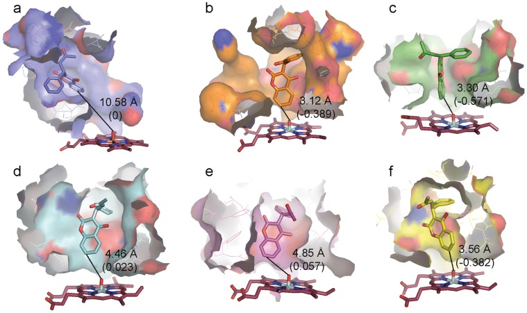
Figure 3. Representative native-like interatomic interactions between (S)-warfarin and CYP2C9 variants.
The total r⋅m⋅r scores were used to discriminate (S)-warfarin-bound conformation from trajectories of each variant. (S)-warfarin poses at different distance from heme center in original 1OG5 (a), re-docked conformation in WT (b). Snapshopts from MD simulations were selected for native-like conformation of WT (c), R144C (d), I359L (e), and L90P (f) by means of knowledge-based scores between hydroxylation site C-7 of (S)-warfarin and molecular oxygen (FeO-C7) as shown in parentheses. FeO-C7 distance of each structure is measured in angstrom. Oxyferryl heme is in ruby red stick while (S)-warfarin is shown in green (WT), light blue (R144C), magenta (I359L), and yellow (L90P) stick. Oxygen and nitrogen are in red and blue, respectively. Carbon is colored based on CYP2C9 variant.

