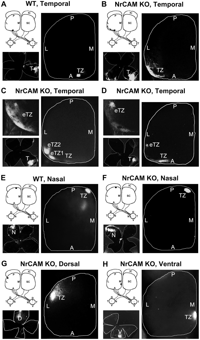
Figure 2. Medial-lateral Targeting Defects of RGC Axons in the Superior Colliculus of NrCAM Null Mice.
A. In WT mice, DiI injection into the temporal retina at P8 labeled a single dense termination zone (TZ) in the anterior SC at P10. B–D. In NrCAM null (KO) mice, DiI injection into the temporal retina labeled multiple laterally displaced ectopic TZs (eTZs) in the SC at P10. E–F. DiI injections into the nasal retina labeled a single TZ in the posterior SC that was normally positioned in WT and NrCAM KO mice at P10. G–H. DiI injections into the dorsal (G) and ventral (H) retina of NrCAM KO mice resulted in normally positioned single TZs in the lateral (G) and medial (H) SC, respectively. The location of DiI injections were shown in retinal flat mounts in the lower left of each image. L, lateral; M, medial; A, anterior; P, posterior; D, dorsal; V, ventral; T, temporal; N, nasal.

