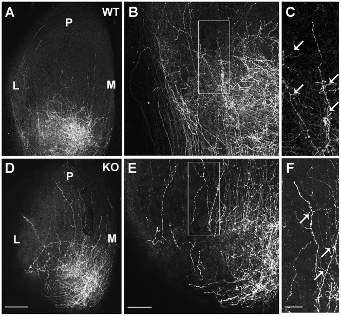
Figure 4. Interstitial Branching of Ventrotemporal RGC axons in WT and NrCAM null SC at P3.
A–C. DiI labeling of VT RGC axons in WT mice showed that most branches from VT axons in the lateral zone of the SC oriented medially to the future SC, as seen in a higher magnification of the boxed area in B (arrows). D–F. DiI labeling of VT axons in NrCAM null mutants (KO) revealed more laterally oriented branches in axons within the lateral zone of the SC, as seen in a higher magnification of the boxed area in E (arrows). M, medial; L, lateral; P, posterior. Scale bar: 200 µm in A,D; 100 µm in B,E; 50 µm in C,F.

