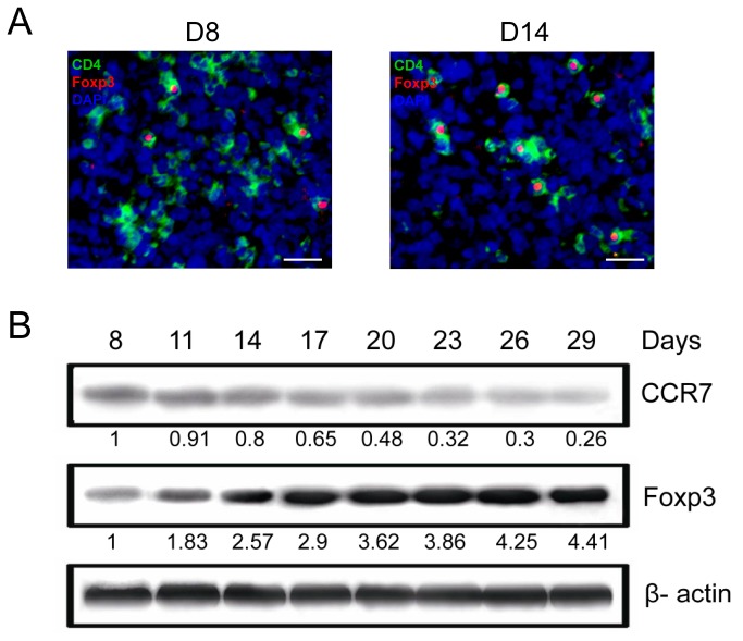
Figure 1. Accumulation of Tregs and different expression patterns of CCR7 and Foxp3 within the progressive tumors.
(A) CD4+ Foxp3+ Tregs were increased in the tumor tissues of early-stage HCC on day 8 and 14 after Hepa1-6 subcutaneous inoculation. Serial 5-µm-thick cryostat sections were double-stained with CD4 (green) and Foxp3 (red) antibodies. Scale bar, 50 µm. (B) CCR7 downregulation and Foxp3 upregulation in the development of HCC from day 8 to 29 were verified by western blot. β-actin was used as an internal control. Protein levels were determined by densitometry analysis and were expressed as ratios to β-actin (below each blot). The ratio obtained from the first lane was set as 1. All data were representative of at least two independent experiments.

