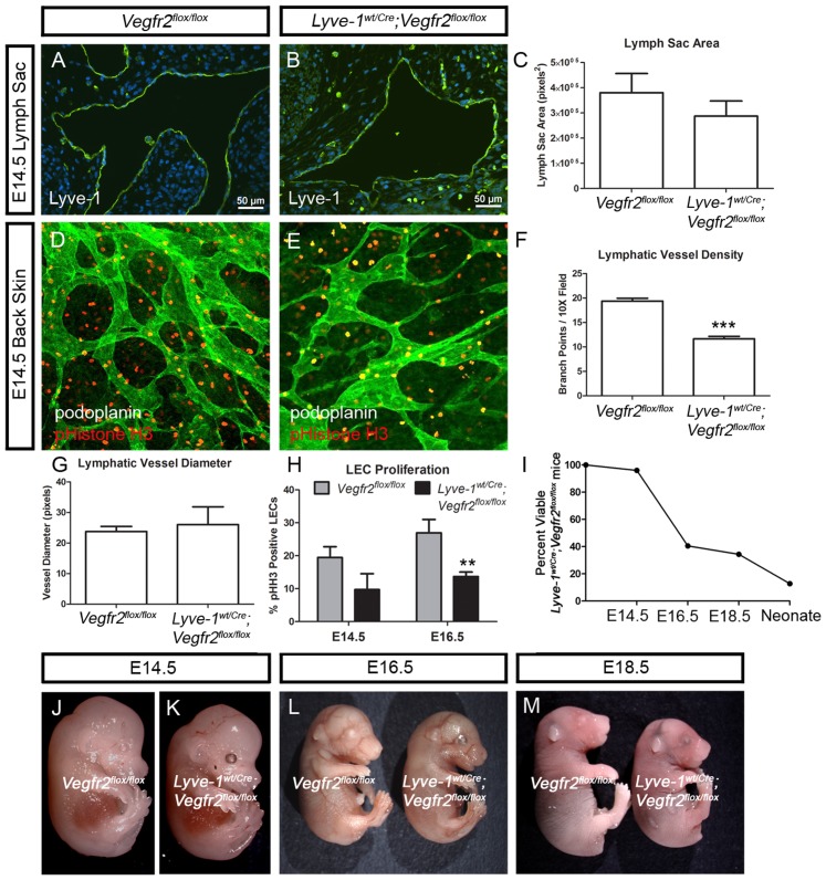
Figure 5. Lyve1wt/Cre;Vegfr2flox/flox embryos exhibit lymphatic defects.
(A,B) Immunofluorescence staining for Lyve-1 showing jugular lymph sacs in E14.5 Vegfr2flox/flox and Lyve-1wt/Cre;Vegfr2flox/flox embryos. (C) Graph showing that lymph sac area is not different between Vegfr2flox/flox (380,110±76,279 pixels2; n = 8) and Lyve-1wt/Cre;Vegfr2flox/flox embryos (287,350±59,931 pixels2; n = 5). (D,E) Images of back skin from E14.5 Vegfr2flox/flox and Lyve-1wt/Cre;Vegfr2flox/flox embryos stained with antibodies against podoplanin (green) and phospho-histone H3 (red). (F) At E14.5, there are significantly fewer lymphatic branch points in Lyve-wt/Cre;Vegfr2flox/flox embryos (11.63±0.5239; n = 3) than in Vegfr2flox/flox embryos (19.37±0.5783; n = 3). (G) At E14.5, lymphatic vessel diameter is not significantly different between Lyve-1wt/Cre;Vegfr2flox/flox (26.00±5.859 pixels; n = 3) and Vegfr2flox/flox embryos (23.75±1.652 pixels; n = 4). (H) Graph showing that there are fewer proliferating lymphatic endothelial cells in Lyve-1wt/Cre;Vegfr2flox/flox embryos than in Vegfr2flox/flox embryos at E14.5 and E16.5. At E14.5, 19.45% of LECs in Vegfr2flox/flox embryos (n = 4) were phospho-Histone H3-positive whereas 9.73% of LECs in Lyve-1wt/Cre;Vegfr2flox/flox embryos (n = 4) were phospho-histone H3-positive. At E16.5, 26.94% of LECs in Vegfr2flox/flox embryos (n = 5) were phospho-Histone H3-positive whereas 13.69% of LECs in Lyve-1wt/Cre;Vegfr2flox/flox embryos (n = 7) were phospho-histone H3-positive. (I) Graph showing the percent of viable Lyve-1wt/Cre;Vegfr2flox/flox mice at different developmental stages. (J-M) Images of non-edematous E14.5 (J,K), E16.5 (L) and E18.5 (M) Vegfr2flox/flox and Lyve-1wt/Cre;Vegfr2flox/flox embryos. ** indicates P || 0.01; *** indicates P || 0.001.

