
Figure 6.
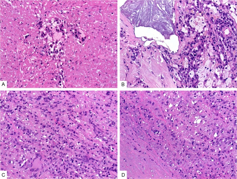
Pattern of elastic fiber damage in lymphoplasmacytic thoracic aortitis. A: A focus of plasma cells associated with elastic fiber loss within the media. B: This example showed dense plasma cell infiltrates abutting a focus of calcification with a few foamy histiocytes. C: This example showed parallel arrays of damaged elastic fibers, note multinucleated giant cells at lower field. D: This case showed subtotal loss of elastic tissue with prominent vacuolation and associated lymphoplasmacytic cells.