
Figure 3.
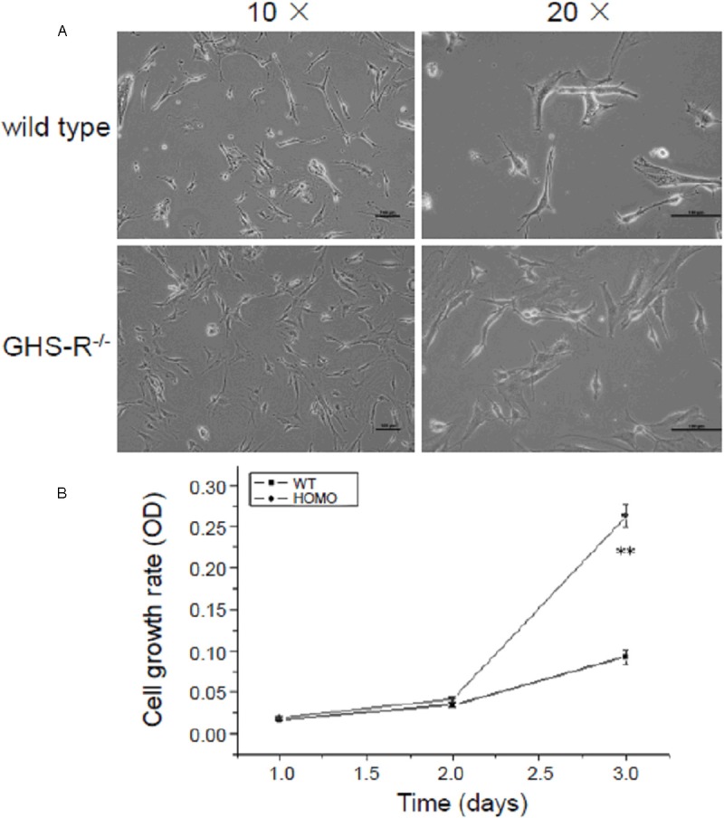
Morphology observation and cell growth analysis of endothelial cells (ECs) from GHS-R-/- mice. A: Morphology observation of in vitro cultivated ECs from GHS-R+/+ and GHS-R-/- mice. Primary ECs were acquired from GHS-R+/+ and GHS-R-/- mice and cultivated in vitro as described in Materials and Methods, and then observed with inverted phase contrast microscopy. ECs from GHS-R+/+ mice were not well adherent to the dish and presented as the round and slender morphology, whereas ECs from GHS-R-/- grew very well and the cytoplasmic membrane was largely spread on the dish. Bar = 100 μm. B: MTT analysis of proliferative rate of ECs from GHS-R-/- mice. The proliferative rate of ECs from GHS-R-/- mice significantly rose up at day 3 compared with that of ECs from wild-type mice. **P < 0.01.