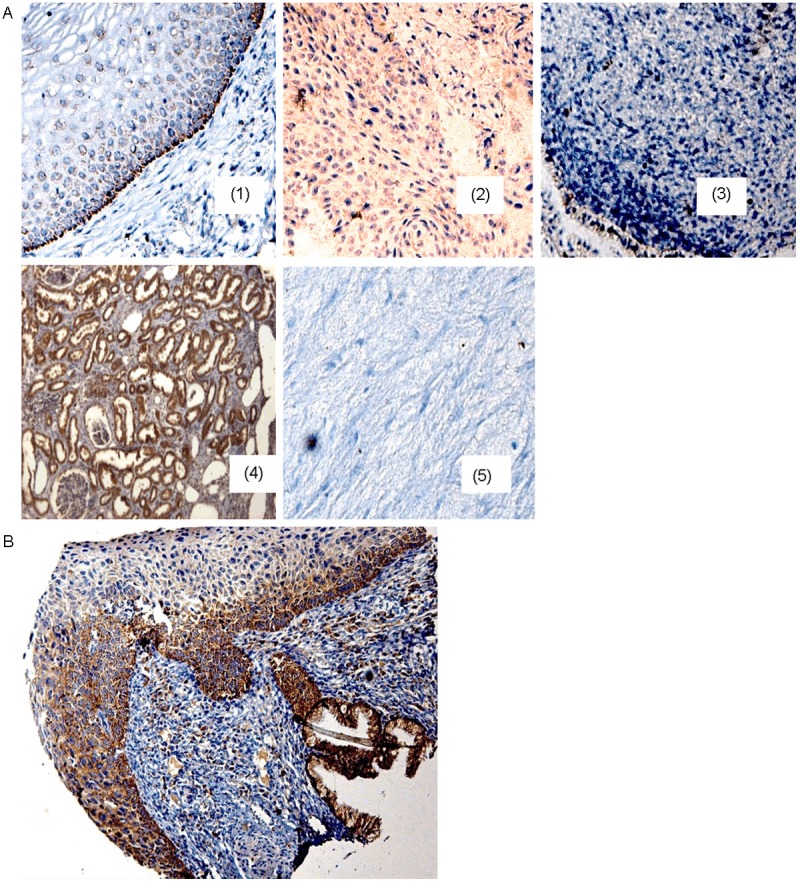
Figure 2.

CRBP1 immunodetection in the uterine cervix samples. A: (1) Cytoplasmic CRBP1 expression is present in cells of the basal layer of normal cervical epithelium (healthy tissue); (2) the immunodetection in the transformed cells of a cervical cancer (CC03) tissue harboring gain of CRBP1 gene. (3) CC samples without gain CRBP1 gene showing negative immunostaining (CC16 sample). A kidney tissue section (4) was used as positive control, while a heart tissue section for negative control (5). B: cervical progression spectrum. The tissue section shows a brownish reaction (positive reaction) in the basal cell layer of the “normal” region, in the high-grade lesion, and also in the invasive region. All tissue sections were hematoxylin counterstained, 200X original amplification.
