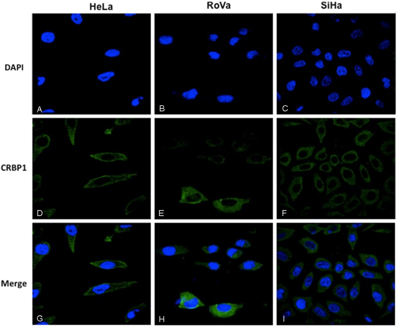
Figure 3.

Immunolocalization of CRBP1 by immunofluorescence in cervical cells. Nuclei were Dapi stained in blue color (A-C). The immunodetection of CRBP1 was observed in green color (D-F). Cytoplasmic immunodetection of CRBP1 in the merge imaging (G-I). 100X original amplification.
