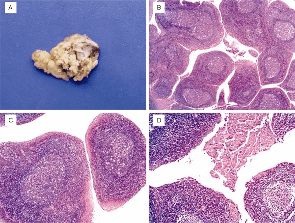
Figure 1.
A: Grossly, the tonsil was occupied by numerous tiny papilliform projections similar to epithelial papillomatosis or lymphoid polyposis on the surface. B: Depicting distinct finger-like projections composed of many phyllodes which contained remarkable follicular lymphoid hyperplasia (H&Ex25). C: The core of the papillary configuration revealed the rich lymphoid tissue with excessive increase of the germinal center and decrease of the follicular cortex (H&Ex100). D: Squamous surface epithelium of the papillae displayed mild hyperplasia with parakeratosis and hyperkeratosis (H&Ex50).

