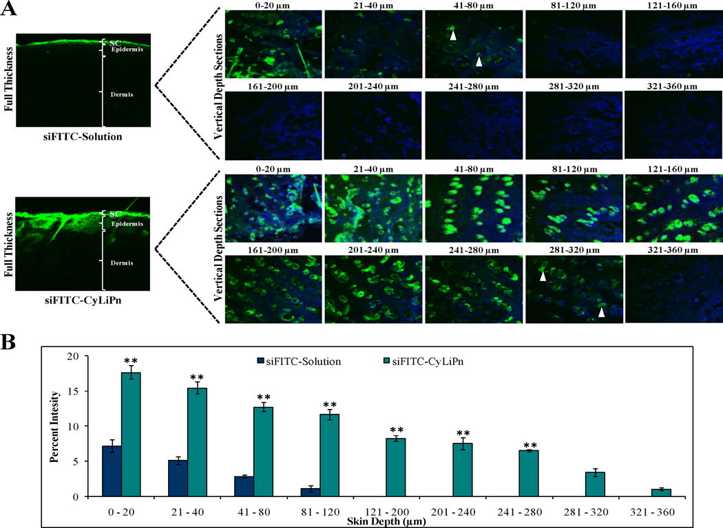
Figure 6.
CLSM images of lateral sections of siFITC permeated skin: After 24 h of in vitro rat skin permeation, skin was collected and lateral skin sections were observed under CLSM. (A) Lateral skin sections of different depths from 0 to 360 µm were observed for skin associated fluorescence. CyLiPn can deliver siFITC (bottom panel) upto 320 µm (white arrows). While, the siFITC-Solution (naked siFITC, Top panel) can penetrate only up to a skin depth of 80 µM (white arrows). The nuclei were stained by Hoechst (blue). (B) The percent fluorescence intensity of the siFITC-Solution and siFITC-CyLiPn permeated skin sections were calculated from confocal images using digital image software. The percent intensity of the images was plotted against the skin depth for different CPPs. Data represent mean ± SD (n=6); significance CyLiPn against solution, **p < 0.001.

